Experience Vision Freedom:
Orbital Fracture Care in Dubai
Home | Services | Comprehensive Orbital Care: Protecting Your Eye's Foundation
Introduction
Imperial Health provides comprehensive care for the orbit, the bony socket that houses the eye. Our specialized team offers advanced diagnostics and treatment for a wide range of orbital conditions, ensuring the health and functionality of this crucial structure. We are dedicated to preserving vision and maintaining the aesthetic integrity of the eye and surrounding tissues.
Our Services
The orbit is a complex area, and our services address a variety of conditions, including:
Orbital Fractures: We provide expert management of orbital fractures, utilizing advanced imaging and surgical techniques to restore the structural integrity of the orbit and prevent long-term complications.
Orbital Tumors: Our team offers diagnosis and treatment for orbital tumors, including both benign and malignant growths. We work closely with other specialists to develop a comprehensive treatment plan tailored to your specific situation.
Thyroid Eye Disease (Graves’ Disease): We provide comprehensive care for thyroid eye disease, managing the ocular manifestations of this autoimmune condition, including proptosis (bulging eyes), eyelid retraction, and double vision.
Inflammatory Orbital Conditions: Our specialists diagnose and treat inflammatory conditions affecting the orbit, such as orbital cellulitis and idiopathic orbital inflammation.
Pediatric Orbital Conditions: We also provide specialized care for children with orbital problems, including congenital anomalies and traumatic injuries.
Why Choose Imperial Health
Key Benefits of Choosing Imperial Health for Orbital Care
Specialized Expertise
Our team comprises experienced ophthalmologists and other specialists with expertise in orbital diseases and surgery.
Advanced Diagonstics
We utilize state-of-the-art imaging and diagnostic techniques to accurately assess your condition and develop an appropriate treatment plan.
Comprehensive Treatment Options
We offer a wide range of treatment options, from medical management to advanced surgical interventions.

Comprehensive Approach
We work closely with other specialists within Imperial Health to provide comprehensive and coordinated care.
Why Choose us
Unique Features and Advantages

Multidisciplinary Approach
We collaborate with other specialists, such as contact lens fitters, to provide comprehensive care.

Focus on Patient Education
We believe in empowering our patients with knowledge about their condition and treatment options.

Commitment to Research
We are actively involved in research to advance the understanding and treatment of keratoconus.
Our testimonials
1000+ Satisfied Patients
Had the best experience at Imperial Healthcare. Had my eyes checked by Dr Vinod, who was very helpful and professional and Kumar was always incredibly friendly.

Had the best experience at Imperial Healthcare. Had my eyes checked by Dr Vinod, who was very helpful and professional and Kumar was always incredibly friendly.

Had the best experience at Imperial Healthcare. Had my eyes checked by Dr Vinod, who was very helpful and professional and Kumar was always incredibly friendly.

Previous
Next
Our team
Meet our doctors
Our administration and support staff all have exceptional people skills
and trained to assist you with all medical enquiries.

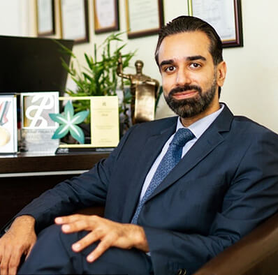
Dr. Vinod Gauba
Medical Director

Dr. Nanis Soltan
MBBS, Msc

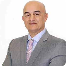
Dr. Samer Hamada
Consultant Ophthalmologist

Dr. Qasim Aref Qasem
Consultant Ophthalmologist
Enquiry Form
Book an Appointment
faqs
Frequently asked questions
SupraLASIK™ is a revolution in laser eye vision correction providing the highest level of safety with superior outcomes
The best feature of SupraLASIK™ is that is in fact NOT LASIK thereby giving it distinct advantages over all the other LASIK procedures. This makes it one of the safest forms of laser eye vision correction available anywhere in the world today.
SupraLASIK™ does not require any cutting or flap therefore it is actually NOT a surgical procedure unlike IntraLASIK or other LASIK procedures. The fact that it does not require any cutting into the cornea confers the many advantages and extremely high safety profile of this procedure.
SupraLASIK™ is performed using the Schwind Amaris excimer laser. This is one of the most sophisticated‚ precise and advanced lasers used in eye surgery in the world.
The procedure usually takes under 60 seconds per eye to perform where there is virtually no contact with the eye itself. The laser simply reshapes the cornea to completely correct shortsightedness (myopia)‚ longsightedness (hyperopia)‚ astigmatism and all other irregularities in the cornea which may affect vision (higher order aberrations).
SupraLASIK™ utilizes all the available advances in laser vision correction optimization such as wavefront customized treatment plans. This means that‚ if required‚ the treatment will not only correct the complete eyeglasses prescription you may have but it also corrects any irregularities you may have in your cornea. This further adds to the precision and refinement of visual outcomes from this procedure.
IntraLASIK‚ UltraLASIK or in fact any other LASIK procedure involves cutting the eye to create a corneal flap. This can be done with a blade or with a laser (eg. IntraLASIK). SupraLASIK™ corrects your vision without any cut into the cornea. This has several advantages since it makes the procedure much safer‚ more comfortable and quicker than other LASIK procedures. Avoiding cutting into the cornea also reduces the impact on corneal nerves created by the procedure. This helps significantly reduce the incidence of dry eye after this procedure which is one of the main complications of other LASIK procedures. Reducing post–treatment dry eye is of great importance not only to ensure long term comfort for the patient but also to help ensure long term stability of vision and reduce the risk of regression.
Once your eye has healed and recovered from the procedure‚ the cornea heals almost completely back to how it was prior to surgery except for the fact that you can now see clearly without glasses. There is almost no evidence of having had the procedure such that even an eye specialist would find it hard to tell unless you informed them beforehand or they specifically check for it with a corneal scan. Also the eye is not any weaker than prior to surgery if it were ever to sustain an injury. In fact it would behave the same way to injury before or after surgery. This is in contrast to other LASIK procedures‚ where direct high force injury to the cornea can displace or even amputate the corneal flap even decades after the original procedure. This is of particular relevance to people playing contact sports or that may be involved in direct contact activities such as members of the armed forces‚ police or certain professional athletes.
The German developed and produced SCHWIND AMARIS laser systems are deemed‚ also by independent experts‚ to be the highest standard among eye lasers for refractive surgery. Above all‚ patients profit from their strong features since treatment with the SCHWIND AMARIS laser systems is not only significantly faster‚ more precise and safer than ever before – it is also considerably more pleasant.
The SCHWIND AMARIS laser systems provide all features of modern laser technology. And they offer even more‚ because in the SCHWIND AMARIS laser systems groundbreaking innovations have been materialized – such as the ablation with two energy levels or the unique eye tracking system. So far‚ these features have not been available in any other device.
Using the Schwind Amaris‚ our ophthalmologist provide advanced and customized laser eye surgery using latest technologies‚ such as the Wave front guided Tortional eye Tracker which can track even the slightest eye movements in the x‚ y and z axis. All these technologies are incorporated into the SupraLASIK™ treatment.
No. Eye drop anesthesia is applied several times before the procedure to numb the cornea however since SupraLASIK™ does not involve any cutting into the eye and almost no touching of your eye during the procedure‚ it is extremely comfortable. The only sensation reported is often that of the saline fluid used at the end of the procedure to keep the cornea wet and clean.
LASEK and PRK all require manual removal of the surface layer of the cornea. In fact both options often use a chemical agent to loosen up the surface layer before scraping it off. SupraLASIK™ does not require any chemicals or scraping of the eye since it uses the safe and painless Schwind Amaris laser to remove this surface layer. Avoiding manual removal of this layer helps to increase safety‚ speed up recovery time and improve overall results and outcomes. Also any potential risk of long term corneal haze is significantly reduced by the use of a special medicine applied at the end of the procedure.
During the first 48 hours after the procedure‚ patients normally experience watery‚ light sensitive eyes. During this period it is best advised to stay at home and avoid prolonged visual activity. After this period the eyes are comfortable but the vision is not stable due to a special protective healing lens that is placed in the eye at the end of surgery. This lens is removed on day 6 after which the vision stabilizes further. The vision initially fluctuates before it becomes crisp and sharp. This recovery period does vary amongst individuals although following the advised post–treatment regime and precautions helps speed up this process. Although the recovery is not as quick as from an uncomplicated LASIK procedure‚ we feel that the initial time investment during the first days after the procedure is well worth the additional benefits or less long term issues.
During the first few days after the procedure‚ you’ll be given special drops to place regularly in your eyes. You will also be clearly advised of all do’s and don′ts. For instance‚ you must avoid rubbing your eyes too much in the first few days. Once the initial healing period is passed‚ the precautions are fairly minimal and you can quite quickly get back to your normal life – except now without glasses or contact lenses!
The survey of our SupraLASIK™ patients tells us that 50% go back to work on day 4 whilst almost 100% go back to work on day 5 after the procedure.
Once healed‚ your eye would react the same way to injury as it would prior to the procedure since SupraLASIK™ does not structurally weaken your eye to injury unlike other types of LASIK.
We are so confident of the safety and accuracy of SupraLASIK™ that we offer a ‘lifetime care guarantee’.
This means that we are so confident that SupraLASIK™ will be a once–in–a–lifetime solution to free you from glasses or contact lenses‚ that if your prescription comes back at any stage in the future‚ we will treat it for free. Clearly we can only offer such a guarantee because we don′t have to perform such re–treatments due to the high accuracy‚ reliability and consistency of SupraLASIK™.
This depends on several factors such as your state of eye lubrication‚ corneal curvature‚ corneal thickness and so on. However since SupraLASIK™ does not cut into the cornea‚ and has a minimal impact on the thickness and strength of the cornea‚ we can safely perform the re–treatment to give spectacle–free vision in suitable candidates.
