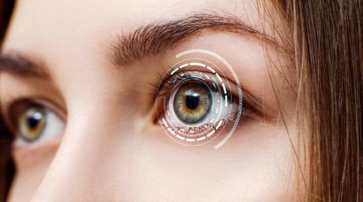
Comprehensive Guide on Retina Functions, Layers, and Signs
The retina is a vital component of the eye, responsible for capturing light and converting it into neural signals that the brain interprets as visual images. Understanding the retina’s functions, its layered structure, and the signs of potential problems is crucial for maintaining eye health. Regular check-ups with a retina specialist in Dubai can help detect and treat retinal issues early, ensuring optimal vision. This guide provides an in-depth look at the retina, its functions, the various layers it comprises, and the common signs indicating retinal problems.
Functions of the Retina
Light Detection and Image Processing
The primary function of the retina is to detect light and convert it into neural signals. This process begins when light enters the eye and strikes the photoreceptor cells—rods and cones—located in the retina. Rods are responsible for vision in low light conditions, while cones detect color and provide sharp central vision. These photoreceptors convert light into electrical signals, which are then processed by other retinal neurons, including bipolar cells and ganglion cells, and transmitted to the brain via the optic nerve. The brain then interprets these signals to create the images we see.
Color Vision
Color vision is another essential function of the retina, primarily mediated by cone cells. There are three types of cones, each sensitive to different wavelengths of light: red, green, and blue. The brain combines the input from these cones to perceive a full spectrum of colors. Any malfunction or deficiency in these cones can lead to color vision deficiencies, commonly referred to as color blindness. Proper functioning of the cones is crucial for activities that rely on color discrimination, such as identifying traffic lights or choosing ripe fruits.
Peripheral and Central Vision
The retina plays a vital role in both peripheral and central vision. Peripheral vision, mediated by rods located around the retina’s edges, allows us to detect movement and navigate our environment. Central vision, provided by cones concentrated in the macula (the central part of the retina), enables us to perform tasks that require detailed vision, such as reading and recognizing faces. A healthy retina ensures that we have a wide field of vision and the ability to focus on fine details.
Layers of the Retina
Photoreceptor Layer
The photoreceptor layer is the outermost layer of the retina, containing rods and cones. Rods are highly sensitive to light and allow us to see in low-light conditions, while cones function best in bright light and are responsible for color vision and detailed central vision. This layer’s proper functioning is essential for capturing light and initiating the process of vision.
Retinal Pigment Epithelium (RPE)
Beneath the photoreceptor layer lies the retinal pigment epithelium (RPE), a layer of pigmented cells that support the photoreceptors. The RPE absorbs excess light, preventing it from scattering within the eye, and plays a crucial role in recycling visual pigments and maintaining photoreceptor health. It also acts as a barrier and regulator of nutrients between the retina and the choroid (the vascular layer of the eye).
Bipolar and Ganglion Cell Layers
The bipolar cell layer receives electrical signals from the photoreceptors and transmits them to the ganglion cells. The ganglion cell layer contains the axons that form the optic nerve, which carries visual information to the brain. These layers are integral in processing and refining the visual signals received from the photoreceptors before they are sent to the brain for interpretation. Proper functioning of these layers ensures that the visual information is accurately conveyed and processed.
Inner and Outer Plexiform Layers
The inner and outer plexiform layers are regions where synapses, or connections, occur between different types of retinal neurons. The outer plexiform layer is where photoreceptors connect with bipolar cells, while the inner plexiform layer is where bipolar cells connect with ganglion cells. These layers facilitate the complex processing of visual information within the retina, enabling the integration and modulation of signals before they are sent to the brain. This synaptic activity is crucial for the accurate perception of visual stimuli.
Signs of Retinal Problems
Blurred or Distorted Vision
Blurred or distorted vision can be an early sign of a retinal problem. Conditions such as macular degeneration, diabetic retinopathy, and retinal vein occlusion can cause the retina to swell or develop abnormal blood vessels, leading to vision changes. Individuals may notice that straight lines appear wavy or that their central vision is blurred. Prompt consultation with an eye care professional is essential for diagnosing and treating the underlying cause of these symptoms.
Floaters and Flashes of Light
The sudden appearance of floaters (tiny spots or strands that drift through the field of vision) or flashes of light can indicate retinal issues such as retinal detachment or posterior vitreous detachment. Floaters occur when small particles in the vitreous gel of the eye cast shadows on the retina, while flashes are caused by the vitreous gel tugging on the retina. If these symptoms occur suddenly and are accompanied by a loss of vision, immediate medical attention is necessary to prevent permanent damage.
Loss of Vision
Partial or complete loss of vision, whether gradual or sudden, is a serious symptom of retinal problems. Conditions like retinal detachment, macular holes, and advanced diabetic retinopathy can lead to significant vision loss if not treated promptly. Peripheral vision loss, known as tunnel vision, can be a sign of glaucoma or retinitis pigmentosa, while central vision loss can indicate macular degeneration. Regular eye examinations can help detect these conditions early, allowing for timely intervention and treatment.
Dark Spots or Shadows
Dark spots or shadows in the visual field, known as scotomas, can be a sign of retinal damage. These may appear as small, dark areas or larger patches that obscure part of the vision. Conditions such as macular degeneration, diabetic retinopathy, and retinal vascular occlusions can cause scotomas. The sudden onset of a shadow or curtain-like effect over the visual field is particularly concerning and can indicate retinal detachment. Immediate medical evaluation is essential to address the underlying cause and prevent further vision loss.
Changes in Color Vision
Changes in color perception, such as colors appearing faded or less vibrant, can indicate retinal diseases like macular degeneration or diabetic retinopathy. These conditions affect the photoreceptor cells responsible for detecting and differentiating colors. Gradual color distortion can impact daily activities that require color discrimination. Recognizing these changes early and seeking professional evaluation can help manage the underlying condition and preserve color vision.
Difficulty Reading
Difficulty reading or seeing fine details can be a sign of macular problems. Macular degeneration, diabetic macular edema, and macular holes can all affect the central vision, making it challenging to read small print or recognize faces. Individuals may notice that they need brighter lighting or magnifying glasses to read comfortably. Early diagnosis and treatment are crucial to slowing the progression of these conditions and maintaining reading ability.
Read More: What are Signs of Retina Problems?
Prevention and Maintenance of Retinal Health
Regular Eye Exams
Regular eye exams are crucial for maintaining retinal health and early detection of retinal disorders. Adults, especially those with risk factors such as diabetes, hypertension, or a family history of retinal diseases, should have comprehensive eye exams annually. These exams can detect early signs of retinal problems, allowing for timely intervention and treatment to prevent vision loss.
Healthy Lifestyle Choices
Adopting a healthy lifestyle can significantly impact retinal health. A balanced diet rich in antioxidants, vitamins, and minerals supports eye health. Regular exercise helps maintain healthy blood circulation, including to the eyes. Avoiding smoking and managing chronic conditions such as diabetes and hypertension are also essential steps in preventing retinal diseases. These proactive measures can reduce the risk of developing conditions like age-related macular degeneration and diabetic retinopathy.
Advances in Retinal Treatments
Anti-VEGF Therapy
Anti-VEGF (vascular endothelial growth factor) therapy has revolutionized the treatment of retinal diseases like wet age-related macular degeneration and diabetic macular edema. VEGF is a protein that promotes abnormal blood vessel growth and leakage in the retina. Anti-VEGF medications, administered through injections into the eye, inhibit this protein, reducing swelling and preventing further vision loss. Regular treatments can stabilize and even improve vision in many patients.
Gene Therapy
Gene therapy offers promising new treatments for inherited retinal diseases such as retinitis pigmentosa and Leber congenital amaurosis. This innovative approach involves delivering healthy copies of genes to replace or repair defective ones in retinal cells. Clinical trials have shown promising results, with some patients experiencing significant improvements in vision. Continued research and development are expected to expand the application of gene therapy in retinal disease treatment, offering hope for those with previously untreatable conditions.
Stem Cell Therapy
Stem cell therapy is another emerging treatment option for retinal diseases. Stem cells have the potential to differentiate into various cell types, including retinal cells. Researchers are exploring ways to use stem cells to replace damaged retinal cells and restore vision. While still in experimental stages, early studies have shown encouraging results, suggesting that stem cell therapy could become a viable treatment for conditions like macular degeneration and retinal degeneration in the future.
Conclusion
The retina’s complex structure and functions are essential for vision. Understanding its roles, recognizing the signs of retinal problems, and seeking prompt treatment can help preserve eye health and prevent vision loss. Advances in diagnostic tools and treatments, such as anti-VEGF therapy, gene therapy, and stem cell therapy, offer new hope for managing retinal diseases. Regular eye exams and healthy lifestyle choices are key to maintaining retinal health. If you experience any symptoms of retinal problems, consult a retina specialist in Dubai to ensure timely and effective care.
Preserve Your Vision with Imperial Healthcare Eye Hospital in Dubai
Imperial Healthcare Eye Hospital in Dubai is dedicated to providing comprehensive care for all your eye health needs. Our team of experienced ophthalmologists specializes in diagnosing and treating a wide range of retinal conditions. Whether you need a routine eye exam or advanced treatment for a complex retinal disorder, our state-of-the-art facilities and personalized care ensure the best possible outcomes. Don’t wait until your vision is compromised—schedule an appointment today and take the first step towards preserving your eyesight. Trust the experts at Imperial Healthcare Eye Hospital, the premier eye hospital in Dubai, to protect and enhance your vision for a brighter future.

